Showing posts with label Evidence based medicine. Show all posts

Friday, 1 March 2019

The Parkinson’s Drug Trial: A Miracle Cure?


I’m sure many of you have seen the programme about a landmark Parkinson’s study, that was aired on the BBC last night (28th February 2019). It is a very well-produced fly-on-the-wall documentary that follows the investigators, inventors and some participants that have pioneered a truly ‘disruptive’ trial. If you haven’t seen it, stop reading and watch now (UK readers only).

The inimitable Tom Isaacs, who was one of the first patients into the study, and who's charity the Cure Parkinson's Trust funded this study.


What made it so unique – after all, there are thousands of drug studies that take place across the UK every day. Usually a study is designed to test one thing – most often a drug. In this study, they didn’t just test if the new drug worked. They had to invent new hardware – that needed to be tested. They had to invent a new kind of brain surgery – that needed to be tested. They had to persuade fairly risk-averse regulatory bodies and ethics panels that doing all three, together, at once was fair, safe and acceptable. Regardless of the outcomes of the study, I can only marvel at the courage and dedication shown by every person involved, from the chief investigator and the whole study team, to each and every participant and their families. Medicine usually progresses by small evolution, this study was a major revolution.


So what did the study show? ***Spoiler alert*** (The second part of the documentary is due to be aired next week. Here I will review the peer-reviewed scientific data that was published in two papers this year)

The one sentence summary from the first paper might, on the surface, appear to indicate defeat, “At the 40-week point, however, we have not shown clinical benefit despite this putamen-wide tissue engagement.” However, despite the headline ‘failure’, there is so much to celebrate.
Firstly, the managed to recruit (in fact, for 42 study ‘spots’, they had over 200 people volunteer). This is no small challenge, especially when there is the very real risk of making people worse off, and asking people with Parkinson’s to travel to Bristol every month for nearly a year.
Secondly, they proved that the hardware and surgery worked, and worked safely. 
Image from BBC.com
Thirdly, and very importantly, they showed that after 40 weeks, there was no change in the dopamine-scan in those who’d had placebo; but in the GDNF group there was a marked change that took the appearances back to those expected at the early stage of Parkinson’s. Bear in mind that on average, the participants had had symptoms of Parkinson’s for over 10 years, and in some for well over 15 years!


Fourthly, at the end of 40 weeks, no patients in the placebo group had movement scores that improved by 10 or more points. However, 9 out of the 17 participants given GDNF improved by between 10 and 16 points. This was not only statistically significant (albeit as a post hoc analysis), but most definitely clinically significant.
So what else can be said? Well, firstly, there was a very strong placebo effect. At one point in the film, the mother of one of the participants says, “To have a huge operation like that, and then have to have a placebo. Is that human? Not really, is it!” The BBC have produced a very good summary on the placebo (and nocebo) effects. The fact that the placebo group did improve so much, means that there is much less of a difference between the two groups. This doesn’t necessarily mean that the GDNF didn’t work, it means that the effect is ‘lost’ in the statistical analysis. 
Furthermore, the numbers in the study were very small, with only 41 patients in total, (21 GDNF, 20 placebo.) This further reduces the chance of finding statistical differences.

A - model showing the novel equipment that was designed and made specifically for this study. B - the port that participants lived with, with which they were 'hooked up' to treatment. C - brain scan showing that the drug delivery worked exactly where it needed to. D - patient sat being infused with treatment via the 'head port'. E - A (not-so) ordinary infusion suite. (image from linked Brain paper)

After the original 40 week study, both groups continued to get another 40 weeks treatment, this time with everyone getting the GDNF. At the end of this phase, there was still no overall group differences from the very beginning to the very end between those who had 80 weeks vs 40 weeks of GDNF treatment, in terms of the movement scores off all treatment. However, those who’d had GDNF all along had fewer increases in their regular medication (to a tune of nearly 5 tablets-worth per day (59mg vs 289mg)). There was also ongoing improvement in the ability to do daily activities in those having GDNF for 80 weeks, and a continued improvement in the proportion of the day when the medication was working well.
There are several other factors that need to be borne in mind. The study cost £3m. Divided over each of the participants amounts to over £73,000 per person. Although this seems like a lot, the actual treatment cost, should this every come to clinical practice would likely be much lower, as it includes all the extra costs associated with running a complex clinical trial. Furthermore, if GDNF is ultimately shown to slow the progression of the condition, if it means that someone maintains their independence and can either carry on working (and paying tax), or stay in their own home independently, rather than take early retirement or move to a nursing home, this really is small change – and when you factor in Quality of Life benefits, must surely be a price worth paying.
Finally (and thank you for bearing with me to this point), all these patients had a diagnosis of Parkinson’s disease for around 8 years. This means that they had the pathology for approaching 20 years. Many were profoundly affected by their symptoms. To use a metaphor I’ve used previously on this blog, it is easier to blow out a match than fight a forest fire. The brain of someone with advanced Parkinson’s is vastly more effected than at the early, or even pre-symptomatic stages. Any restorative attempts are far more likely to work if you can catch it early and intervene early. This is the overwhelming conclusion from treating infections and the whole concept of cancer treatment. 
Hopefully, you and your friends will help us reach that goal with Parkinson’s. You can do your bit by helping us to identify the very earliest phase of Parkinson’s: www.predictpd.com. Taking part involves 25 minutes in front of your own computer at home, and certainly doesn’t pose any of the risks that this study did!
As a final note, I would like to say that this programme reminded me how much of a privilege it is to look after people with Parkinson’s in the clinics of the Royal Free and Luton & Dunstable Hospital; how much of a privilege it is to be at the forefront of Parkinson’s research, funded by Parkinson’s UK, and how much this motivates me while training for the London Landmarks half-marathon and Virgin Money London Marathon.
RNR 

Thursday, 1 November 2018

PLAIN ENGLISH: Does removal of the appendix impact the risk of Parkinson's Disease?

Multiple large news organisations yesterday were reporting the results of a study showing that people who have had appendectomies (surgical removal of the appendix) have a lower chance of developing Parkinson's disease. Its a neat paper which links large amounts of data from medical databases and histology results (looking at tissue under the microscope) on a much smaller number of patients.

First they looked at the Swedish National Patient Registry, a database of 1,698,000 people followed up for 52 years, and identified anyone who had a history of an appendectomy (551,647 people). They then matched these patients to two 'control' patients who had not had appendectomies but were similar in age, sex and geographical to the original patients. They interestingly found that the chance of having Parkinson's disease was 19.3% lower in the participants with appendectomies than the 'control' participants who had not had appendectomies. They also found that the participants who had appendectomies and developed Parkinson's on average developed Parkinson's 1.6 years later than the Parkinson's patients who still had their appendix. Interestingly this effect was not observed in participants who had known genetic mutations causing their Parkinson's suggesting that if the appendix has any effect on the development of Parkinson's it is only in patients whose Parkinson's has an 'environmental' trigger or cause.

This part of the study was not novel or new and previous similar studies have been done and found no association between appendectomy and PD. In a Canadian study they actually found a higher rate of PD in patients who had appendectomies in the past 5 years. So despite the results of this study being very interesting they are not backed up by previous studies.

The novel part of this study is the histological analysis where the researchers analysed the 48 peoples appendixes under the microscope. These participants did not have PD. The appendixes were analysed under the microscope for clumps of protein which have stuck together that we call 'aggregates of α-syneuclein'. These are the key feature of PD that can be found in PD patients brains at autopsy. In other words the misfolded protein that causes PD when it is seen in the brain can be seen in young, healthy peoples appendix. These misfolded proteins were detected in both young and old people.

There are some problems with this study; first is that in large databases it is very difficult to know the accuracy of the Parkinson's diagnosis and previous studies investigating the reliability of this found only a 70.8% chance that it is reliable. They also only examined the appendix under the microscope and did not check any other tissues to compare.

This study does provide some food for thought; at the end of the paper the authors hypothesise that the appendix is the source of the misfolded protein that then spreads through the gut and up the vagus nerve to the brain thereby causing Parkinson's disease. This gut to brain model of the development of Parkinson's disease is often discussed and is a popular research topic at the moment. However the results of this study should still be taken with a pinch of salt because there are a number of large scale similar studies that precede this study that do not come to the same conclusion, that doesn't stop it being interesting though!

Friday, 31 August 2018

Raising a glass to Parkinson's?

Lifestyle factors have been studied at length in relation to Parkinson's Disease and there is good evidence that having ever smoked is associated with reduced risk of the disease. Alcohol drinking has also been linked to reduced risk of disease, and multiple studies have looked at the association. The difficulty with drawing robust conclusions in this area relates to the design of the studies - many of the studies are case control, taking people with and without the disease and asking them to report whether and how much they drink now and in the past. This kind of study suffers from problems with bias - if you have been diagnosed with the disease you are more likely to have attended hospital, to be the kind of person who worries about their health and so makes more health-conscious choices, you may recall things differently in light of your diagnosis and you are likely to have made different choices subsequent to diagnosis.

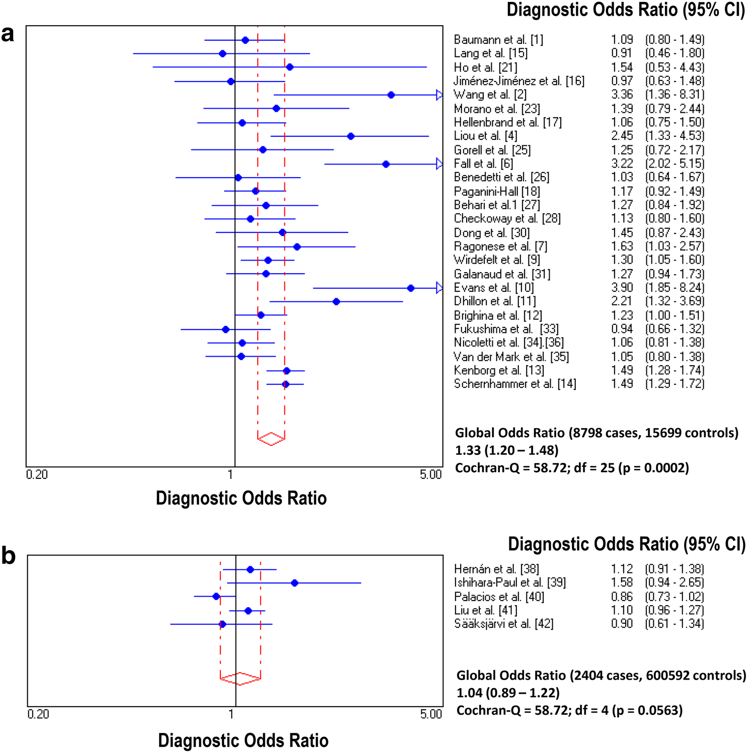
Longitudinal studies of populations who are healthy at baseline are less likely to suffer from these biases as reporting of alcohol consumption is made prior to subsequent diagnosis. This problem with investigating causation in medicine is illustrated nicely by a study published in the Journal of Neurology by a Spanish team. They looked at 31 studies in the field and broke down the analysis into different types of study as well as looking seperately at men and women and different ways of classifying alcohol consumption.

When looking at the case-control studies, as in previous studies, they found higher numbers of 'never-drinkers' in the group with Parkinson's. However, when looking at longitudinal studies, there was no difference in having ever drunk alcohol between people who went on to develop Parkinson's and people who didn't. However, there were far more case-control studies in the article, giving more power to detect these kind of effects, as  you can see in the diagram below.


https://media.springernature.com/original/springer-static/image/art%3A10.1007%2Fs00415-018-9032-3/MediaObjects/415_2018_9032_Fig2_HTML.png
Pooled odds ratios comparing "never drinking" to "ever drinking"alcohol in a)case control and b)prospective longitudinal studies, showing higher likelihood of Parkinson's disease in those who never drank in case control but not longitudinal studies. From https://link.springer.com/article/10.1007/s00415-018-9032-3 
All in all, alcohol consumption remains an interesting part of the puzzle of Parkinson's causation, especially given possible genetic links the authors discuss between variants of the alcohol dehydrogenase gene which cause adverse effects of alcohol and dopaminergic function. In the PREDICT-PD study we are collecting data on alcohol use in all of our participants and will continue to study these links.

-Anna

J Neurol. 2018 Aug 28. doi: 10.1007/s00415-018-9032-3. [Epub ahead of print]
Alcohol consumption and risk for Parkinson's disease: a systematic review and meta-analysis.
Jiménez-Jiménez FJ, Alonso-Navarro H, García-Martín E, Agúndez JAG
The possibility that alcohol consumption should be considered as a "protective factor" for Parkinson's disease (PD) has been suggested by several case-control studies. However, other case-control studies and data from prospective longitudinal cohort studies have been inconclusive. We carried out a systematic review which included all the eligible studies published on PD risk related with alcohol consumption, and conducted a meta-analysis according to the preferred reporting items for systematic reviews and meta-analyses (PRISMA) guidelines. The systematic review was performed using two databases, and the meta-analysis of the eligible studies with the software Meta-Disc1.1.1. Heterogeneity between studies was tested with the Q-statistic. The meta-analysis included 26 eligible retrospective case-control studies (8798 PD patients, 15,699 controls) and 5 prospective longitudinal cohort studies (2404 PD patients, 600,592 controls) on alcohol consumption and PD. In retrospective case-control studies the frequency of PD patients never drinkers was higher and the frequency of heavy + moderate drinkers was lower [diagnostic OR (95% CI) 1.33(1.20-1.48) and 0.74(0.64-0.85)], respectively, when compared to healthy controls. In contrast, in prospective studies, the differences were not significant with the exception of a trend towards a higher frequency of non-drinkers in PD women and a significantly lower frequency of moderate + heavy drinkers in PD men in those studies which stratified data by gender. The present meta-analysis suggests an inverse association between alcohol consumption and PD, which is supported by the results of case-control studies but not clearly by prospective ones.


Monday, 23 July 2018

Remedies from the natural world

An article caught my eye this week as several comments received on this blog and in clinic relate to ‘natural’ remedies for Parkinson’s. To provide some background, my therapeutic philosophy is a common one: I will only prescribe or endorse treatments that have gone through robust, unbiased, reproducible testing, and where the benefits outweight the risks. I also believe that there are undoubtedly treatments that have the power to radically alter diseases that are under our noses in the natural world, and once they are identified, purified and tested, will revolutionise medicine. Exenatide is a perfect example: originally derived from the saliva of the Gila monster, this is now a powerful drug against diabetes and may even have the ability to halt or slow Parkinson’s. 

This article looks at whether blackberry juice can reduce levodopa-induced dyskinesia. The first thing to point out was that they didn’t test it in humans with Parkinson’s disease, this experiment involved the MPTP mouse model of Parkinson’s. Mice were given the same amount of levodopa and then different groups were given increasing doses of blackberry juice. The good news is that there was some benefit on the extra movements associated with ‘dopamine overdose’. However, this was mainly seen in the 15mL/kg group. To put this into a human context (assuming that it works as well in humans with Parkinson’s as mice with MPTP induced parkinsonism, which is a very  big assumption, a human would have to drink over a litre of pure blackberry juice per day to have less dyskinesia. 

Science is a journey of increments. These positive results may yield a useful weapon for people with Parkinson’s, but plenty of work needs to be done. I highly recommend blackberries, especially ones forraged on a nice walk in the woods, but I wouldn’t suggest they’re going to make your Parkinson’s better just yet.

RNR

Effects of blackberry (Morus nigra) fruit juice on levodopa-induced dyskinesia in a mice model of Parkinson's disease.
J Exp Pharmacol. 2018 Jul 4;10:29-35. doi: 10.2147/JEP.S161782. eCollection 2018.


Abstract
BACKGROUND AND OBJECTIVE: 
Levodopa-induced dyskinesia (LID) is a movement disorder that occurs due to levodopa consumption for a long period to attenuate Parkinsonism. Plants have been the basis for medical treatments in human history and still widely practiced. Blackberry (Morus nigra) is one of the fruits rich in anthocyanin. The present study examined the effect of blackberry fruit juice on LID in 1-methyl-4-phenyl-1,2,3,6-tetrahydropyridine (MPTP)-induced Parkinson's disease in mice.
MATERIALS AND METHODS: 
In this study, 42 male mice were used, which were divided into six groups equally: one control group and five groups receiving MPTP injection. After confirmation of Parkinsonism in MPTP groups, one group was preserved without treatment and four other groups were treated with levodopa (50 mg/kg ip). After the onset of LID (2 weeks), one group was kept without additional treatment and three other groups were treated with three different doses of blackberry fruit juice (5, 10, and 15 mL/kg) with levodopa orally for 7 days. Abnormal involuntary movement scale (AIMS) and cylinder behavioral test were carried out according to the schedule. The collected data were analyzed using the SPSS software with the significant level of P<0.05.
RESULTS: 
Parkinson's disease was confirmed with AIMS test on the fourth day after MPTP injection. The onset of LID was observed after 2 weeks of levodopa treatment using both behavioral tests. The result of administration of M. nigra fruit juice for 1 week showed that this addition is useful in hindering LID. These effects were more pronounced at doses 10 and 15 mL/kg with nearly the same results on attenuating AIMS. Low dose of the fruit juice does not seem to affect LID significantly.
CONCLUSION: 
M. nigra fruit juice is effective to attenuate LID in an MPTP-induced Parkinson mice model.

Wednesday, 27 June 2018

Is the horizon getting closer ?


Week after week we talk about the need for a disease modifying drug in Parkinson’s, and week after week we hope to reassure you, dear reader, (as well as ourselves) that this Holy Grail is getting ever closer. 

This week marks the publication of a study that may mark a turning point in this journey. Prothena Pharmeceuticals have run a study looking at the safety and tolerability of a new compound (excitingly called PRX002/RG7935). This is a compound that sticks to the toxic protein that is understood to cause the brain cell loss in Parkinson’s – α-synuclein. Once it sticks to this substance, it ‘flags’ the body’s immune system to come and clear up the mess. This, we think, reduces the amount of the α-synuclein that is floating around and causing damage. 

In this study 80 participants with mild-moderate Parkinson’s were given one of six different doses of this medicine or salt-water (placebo) through a ‘drip’. They attended once a month and the infusion lasted 1-2 hours. Neither the participants nor the investigators knew how much (if any) of the drug they were getting. They received three infusions in total.

I must stress that this study wasn’t designed to see if this did anything at all for their Parkinson’s. The study was too small, and too short for any changes to be detected. The primary purpose of this study was to evalute whether the medicine was safe and tolderable, with secondary objectives of studying what happens to the drug when it is in the body.

Overall, of the 55 participants that had any strength of the medicine, 50 completed the study, and 5 did not manage all 24 weeks of the study. 1 stopped because their Parkinson’s worsened, 1 decided to withdraw (reason not stated), 2 (both in the highest dose group) stopped because of side effects, and 1 also in the highest group withdrew. I also note that 3 out of 25 getting placebo also withdrew.

This study is a vital step. After extensive laboratory testing, a compound like this will be given to a very small number of totally healthy volunteers to ensure it is safe in humans. Then it will be given to a small number of volunteers with the condition to make sure it is safe in the ‘target’ population and to determine a range of safe doses, which is this study. Then it will be given to a moderately large sample to see if there are any suggestions of positive effects and to refine the ideal dose, and finally it will undergo a large study of affected individuals where the investigators (and cruicially the funders) hope to find that it has a true benefit.

All the participants in this study had had Parkinson’s for around 3 years (although this was as much as 15 years) and was moderately advanced. We hope that in future studies, the investigators take people with the earliest possible disease duration and learn lessions from the Alzheimer’s journey. 

This compound has been being studied in the earliest stages of the research process for around 10 years. The next stage of the path for this compound, the PASADENA trial, started almost exactly 1 year ago, and will be running until February 2021. I would expect results to come out towards the end of 2021, and then (hopefully) a phase 3 study will start which will take several more years to run and analyse. If all these steps are successful, we may be in a position to prescribe this medication in around 7-10 years from now.

RNR


Safety and Tolerability of Multiple Ascending Doses of PRX002/RG7935, an Anti-α-Synuclein Monoclonal Antibody, in Patients With Parkinson Disease: A Randomized Clinical Trial.

JAMA Neurol. Published online June 18, 2018. doi:10.1001/jamaneurol.2018.1487

Joseph Jankovic, MD; Ira Goodman, MD; Beth Safirstein, MD; Tonya K. Marmon, DrPH; Dale B. Schenk, PhD; Martin Koller, MD, MPH; Wagner Zago, PhD; Daniel K. Ness, DVM, PhD; Sue G. Griffith, MD, PhD, MRCP; Michael Grundman, MD, MPH; Jay Soto, BS; Susanne Ostrowitzki, MD, PhD;Frank G. Boess, PhD; Meret Martin-Facklam, PhD; Joseph F. Quinn, MD; Stuart H. Isaacson, MD; Omid Omidvar, MD; Aaron Ellenbogen, DO;Gene G. Kinney, PhD
Importance:Aggregated α-synuclein is believed to be central to the pathogenesis of Parkinson disease (PD). PRX002/RG7935 (PRX002) is a humanized monoclonal antibody designed to target aggregated forms of α-synuclein, thereby inhibiting neuron-to-neuron transfer of presumed pathogenic forms of α-synuclein, potentially resulting in neuronal protection and slowing disease progression.

Objective:To evaluate the safety and tolerability of multiple intravenous infusions of PRX002 in patients with idiopathic PD.

Design, Setting, and Participants:Multicenter, randomized, double-blind, placebo-controlled, multiple ascending-dose trial at 8 US study centers from July 2014 to September 2016. Eligible participants were aged 40 to 80 years with mild to moderate idiopathic PD (Hoehn and Yahr stages 1-3).

Interventions:Participants were enrolled into 6 ascending-dose cohorts and randomly assigned to receive PRX002 (0.3 mg/kg, 1.0 mg/kg, 3.0 mg/kg, 10 mg/kg, 30 mg/kg, or 60 mg/kg) or placebo. Participants received 3 intravenous infusions every 4 weeks of PRX002 or placebo and were monitored during a 24-week observational period.

Main Outcomes and Measures:Safety and tolerability assessments included physical and neurological examinations, laboratory tests, vital signs, and adverse events. Pharmacokinetic parameters included maximum PRX002 concentration, area under the curve, and half-life.

Results:Of the 80 participants, most were white (97.5%; n = 78) and male (80%; n = 64); median (SD) age was 58 (8.4) years. PRX002 was generally safe and well tolerated; no serious or severe PRX002-related treatment-emergent adverse events (TEAEs) were reported. The TEAEs experienced by at least 5% of patients receiving PRX002, irrespective of relatedness to study drug, were constipation (9.1%; n = 5), infusion reaction (7.3%; n = 4), diarrhea (5.5%; n = 3), headache (5.5%; n = 3), peripheral edema (5.5%; n = 3), post-lumbar puncture syndrome (5.5%; n = 3), and upper respiratory tract infection (5.5%; n = 3). No antidrug antibodies were detected. Serum PRX002 levels increased in an approximately dose-proportional manner; mean terminal elimination half-life was similar across all doses (10.2 days). Rapid dose- and time-dependent mean reductions from baseline vs placebo in free serum α-synuclein levels of up to 97% were seen after a single infusion at the highest dose (F78,284 = 1.66; P = .002), with similar reductions after 2 additional infusions. Mean cerebrospinal fluid PRX002 concentration increased with PRX002 dose and was approximately 0.3% relative to serum across all dose cohorts.

Conclusions and Relevance:Single and multiple doses of PRX002 were generally safe and well tolerated and resulted in robust binding of peripheral α-synuclein and dose-dependent increases of PRX002 in cerebrospinal fluid, reaching cerebrospinal fluid concentrations that may be expected to engage extracellular aggregated α-synuclein in the brain. Findings support the design of an ongoing phase 2 clinical study (NCT03100149).

Trial Registration:ClinicalTrials.gov Identifier: NCT02157714.

Tuesday, 30 January 2018

Seeing the future

We are proud to announce another research paper from the Predict-PD stable. We have published our data from the imaging substudy of the first phase of our research.

50 individuals who had gone through the website had a scan showing dopamine-releasing areas in the brain (known as a DAT-SPECT or DATSCAN) and transcranial ultrasound. The DATSCAN is the closest we have to a scan that gives proof of Parkinson’s in the brain. Ultrasound shows an area in the region most affected by Parkinson’s that is brighter in people with Parkinson’s compared to healthy people.

Our findings show that the reductions in the dopamine scan were correlated with risk scores, worse movement scores both on clinical assessment and using the keyboard tapping test, and worse sense of smell. The ultrasound scan also correlated with risk score and motor score on clinical assessment.

These findings show that the risk score calculated from the responses on the website are associated with changes on brain scans that are associated with established Parkinson’s. This is exciting news, not just for us, but it has established that there are hard changes in the brains of those at the highest risk that are very likely to be the earliest signs of Parkinson’s. These scans may well be used to prove eligibility for people to be included  in future drug trials to slow, stop or prevent the diagnosis.

What are the next steps? I’ll be repeating this on a larger sample, and not just using DATSCAN and ultrasound but MRI too, which will give a highly detailed map of the strucutre of the brain. I look forward to sharing these results with you in the next few years.

RNR


Dopamine reuptake transporter–single-photon emission computed tomography and transcranial sonography as imaging markers of prediagnostic Parkinson's disease
Authors
Alastair J. Noyce MRCP, PhD, John Dickson PhD, Richard N. Rees MRCP, Jonathan P. Bestwick MSc, Ioannis U. Isaias MD, PhD, Marios Politis MD, PhD, Gavin Giovannoni FRCP, PhD, Thomas T. Warner FRCP, PhD, Andrew J. Lees FRCP, MD, Anette Schrag FRCP, PhD

  • First published: 30 January 2018


ABSTRACT
Objective: The objective of this study was to examine whether prediagnostic features of Parkinson's disease (PD) were associated with changes in dopamine reuptake transporter–single-photon emission computed tomography and transcranial sonography.
Methods: Prediagnostic features of PD (risk estimates, University of Pennsylvania Smell Identification Test, Rapid Eye Movement Sleep Behavior Disorder Screening Questionnaire, and finger-tapping scores) were assessed in a large cohort of older U.K. residents. A total of 46 participants were included in analyses of prediagnostic features and MDS-UPDRS scores with the striatal binding ratio on dopamine reuptake transporter–single-photon emission computed tomography and nigral hyperechogenicity on transcranial sonography.
Results: The striatal binding ratio was associated with PD risk estimates (P = .040), University of Pennsylvania Smell Identification Test (P = .002), Rapid Eye Movement Sleep Behavior Disorder Screening Questionnaire scores (P = .024), tapping speed (P = .024), and MDS-UPDRS motor scores (P = .009). Remotely collected assessments explained 26% of variation in the striatal binding ratio. The inclusion of MDS-UPDRS motor scores did not explain additional variance. The size of the nigral echogenic area on transcranial sonography was associated with risk estimates (P < .001) and MDS-UPDRS scores (P = .03) only.
Conclusions: The dopamine reuptake transporter–single-photon emission computed tomography results correlated with motor and nonmotor features of prediagnostic PD, supporting its potential use as a marker in the prodromal phase of PD. Transcranial sonography results also correlated with risk scores and motor signs.


Thursday, 25 January 2018

On the road to nowhere


This week brings the publication of the (much-anticipated) EXPEDITION 3 study. This reports the results of a very large drug trial of a drug that is hoped to reduce the progression of Alzheimer’s disease. As you might be able to tell by the name, it has been preceded by two previous similar studies.

What is unique about this study? Firstly, the people involved all had ‘early’ Alzheimer’s disease. Secondly, participants had to have ‘evidence’ of Alzheimers pathology – this is key because in the previous studies although all the participants had dementia, a significant number ended up having dementia of another cause.

What are the other notable aspects of the design? The scales they used assessed ‘everyday’ performance (as best as a validated scale can), rather than an abstract cognitive assessment; the study compared cognitive performace after 18 months of treatment, which is a reasonable time given the tradeoff of the cost of a trial such as this, and the rate of progression of the disease.

What are the results? Unfortunately, there was no significant difference when comparing the two groups after 18 months.

There are many lessons for researchers in the Parkinson’s world to learn from this study. A potential reason for failure is that even at the stage of ‘mild’ dementia, there may be too much ‘toxic protein’ in the brain for the process to be reversed, or slowed. To overcome this would require recruiting people with evidence of the disease, but without symptoms. Fortunately, this is exactly whom we hope to identify in PREDICT-PD. Another potential reason for the failure of this drug in three trials is that with solenizumab, they’re barking up the wrong tree. The drug has been specifically designed to reduce the amount of abnormal amyloid protein in the brain. However, although there is strong evidence for the Amyloid Hypothesis, the lack of efficacy of anti-amyloid treatments has brought many in the field to challenge the concept. In Parkinson’s there is less doubt over the Synuclein Hypothesis, and the challenge remains to find a compound that can reduce it, or stop it accumulating and causing brain destruction.

RNR

Trial of Solanezumab for Mild Dementia Due to Alzheimer’s Disease


Abstract
BACKGROUND
Alzheimer’s disease is characterized by amyloid-beta (Aβ) plaques and neurofibrillary tangles. The humanized monoclonal antibody solanezumab was designed to increase the clearance from the brain of soluble Aβ, peptides that may lead to toxic effects in the synapses and precede the deposition of fibrillary amyloid.
METHODS
We conducted a double-blind, placebo-controlled, phase 3 trial involving patients with mild dementia due to Alzheimer’s disease, defined as a Mini–Mental State Examination (MMSE) score of 20 to 26 (on a scale from 0 to 30, with higher scores indicating better cognition) and with amyloid deposition shown by means of florbetapir positron-emission tomography or Aβ1-42 measurements in cerebrospinal fluid. Patients were randomly assigned to receive solanezumab at a dose of 400 mg or placebo intravenously every 4 weeks for 76 weeks. The primary outcome was the change from baseline to week 80 in the score on the 14-item cognitive subscale of the Alzheimer’s Disease Assessment Scale (ADAS-cog14; scores range from 0 to 90, with higher scores indicating greater cognitive impairment).
RESULTS
A total of 2129 patients were enrolled, of whom 1057 were assigned to receive solanezumab and 1072 to receive placebo. The mean change from baseline in the ADAS-cog14 score was 6.65 in the solanezumab group and 7.44 in the placebo group, with no significant between-group difference at week 80 (difference, −0.80; 95% confidence interval, −1.73 to 0.14; P=0.10). As a result of the failure to reach significance with regard to the primary outcome in the prespecified hierarchical analysis, the secondary outcomes were considered to be descriptive and are reported without significance testing. The change from baseline in the MMSE score was −3.17 in the solanezumab group and −3.66 in the placebo group. Adverse cerebral edema or effusion lesions that were observed on magnetic resonance imaging after randomization occurred in 1 patient in the solanezumab group and in 2 in the placebo group.
CONCLUSIONS
Solanezumab at a dose of 400 mg administered every 4 weeks in patients with mild Alzheimer’s disease did not significantly affect cognitive decline. (Funded by Eli Lilly; EXPEDITION3 ClinicalTrials.gov number, NCT01900665.)


Mild Parkinsonian Signs in a Community Population

One question that many of the PREDICT-PD participants ask me is “I am slower than I used to be, does it mean that I am getting Parkinson’...