Showing posts with label imaging. Show all posts

Wednesday, 23 January 2019

A scan to predict dementia?

We know that Parkinson's is caused by damage to a small area at the base of the brain known as the Substantia Nigra, which contains cells that produce the chemical messenger dopamine. However, this is not the entire picture, and we believe that other brain structures and chemicals may be involved in causing the variety of different symptoms we see in patients. 

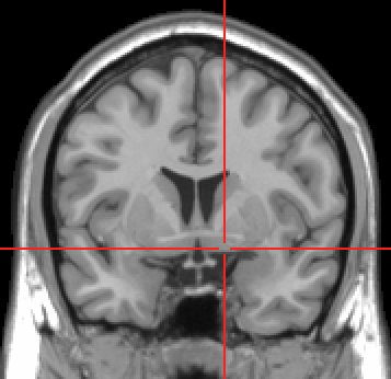
One of the key questions for us in predicting what might happen to patients in the future is whether there is damage that can be found at an early stage, before patients start having problems.  A recent study used hundreds of MRI scans from people with Parkinson's to look at another small structure deep in the brain, named the Nucleus Basalis of Meynert. This is shown in the MRI image below.

https://upload.wikimedia.org/wikipedia/commons/3/36/Substantia_innominata_MRI.PNG
Nucleus Basalis of Meynert
When this team looked at many different brain structures, it was only the size of this part of the brain that could predict whether patients would go on to develop problems with thinking and processing information. This ties in with other work showing this part of the brain plays a key role in different types of dementia as well as our knowledge that a drug which targets the main chemical found in this part of the brain, acetylcholine, is effective in Parkinson's Disease with dementia. 

As researchers continue to put together these kinds of findings, we can develop increasingly accurate ways to predict disease and ultimately ensure that the right people have access to treatments at the earliest stages. 

If you're interested in reading more, here is the link to the article https://academic.oup.com/brain/article/141/5/1501/4944714 

-Anna


Wednesday, 14 November 2018

Magnetic resonance T1w/T2w ratio: A parsimonious marker for Parkinson's disease

I love it when something simple yet really effective...

We are in an era when imaging and laboratory investigations are ever more complex and expensive, and here we have a wonderful study showing that a very simple, cheap and easily-obtained ratio from an MRI scan can be used to accurately diagnose Parkinson's. Importantly the researchers were not satisfied with showing this once and went on to demonstrate similarly impressive results in a separate patient and control group.

Now it is important for other researchers to replicate these findings and to work how far back they go... could it be used to detect people in the pre-symptomatic/pre-diagnostic phase??

Du G, Lewis MM, Sica C, Kong L, Huang X
Ann Neurol 2018 Nov 8


Objective
Newer MRI techniques have shown promise in capturing early Parkinson's disease (PD)‐related changes in the substantia nigra pars compacta (SNc), the key pathological loci. Their translational value, however, is hindered by technical complexity and inconsistent results.

Methods
A novel yet simple MRI contrast, the T1w/T2w ratio, was used to study 76 PD patients and 70 controls. The T1w/T2w ratio maps were analyzed using both voxel‐based and region‐of‐interest approaches in normalized space. The sensitivity and specificity of the SN T1w/T2w ratio in discriminating between PD and controls also were assessed. In addition, its diagnostic performance was tested in a subgroup of PD patients with disease duration ≤2 years (PDE). A second independent cohort of 73 PD and 49 controls was used for validation.

Results
Compared to controls, PD patients showed a higher T1w/T2w ratio in both the right (cluster size=164 mm3, p<0.0001) and left (cluster size=213 mm3, p<0.0001) midbrain that was located ventrolateral to the red nucleus and corresponded to the SNc. The region‐of‐interest approach confirmed the group difference in the SNc T1w/T2w ratio between PD and controls (p<0.0001). The SNc T1w/T2w ratio had high sensitivity (0.908) and specificity (0.80) to separate PD and controls (AUC=0.926), even for PDE patients (AUC=0.901, sensitivity=0.857, specificity=0.857). These results were validated in the second cohort.

Interpretation
The T1w/T2w ratio can detect PD‐related changes in the SN and may be used as a novel, parsimonious in vivo biomarker for the disease, particularly early stage patients, with high translational value for clinical practice and research.

Thursday, 15 March 2018

Treat the person not the scan


When learning clinical medicine, (junior) doctors can often show an over-reliance on blood tests and scans, rather than focussing on the patient and their holistic care. This can lead in turn to more intensive investigation, which is both costly and incurs extra risk, as well as in turn creating more ‘abnormal results’.
An important report has been published in pre-symptomatic Alzheimer’s this week, in the journal Lancet Neurology. The research team at the Salpêtrière Hospital in Paris studied over 300 older adults with subjective, but no objective, memory complaints. The participants completed detailed neuropsychological and cognitive assessment, structural and functional brain scans (MRI and PET), brain-wave tests (EEG), genetic testing and lumbar punctures. These tests were repeated every 6-12 months for over 2 years. They set out to see if they could identify particular markers that would identify who would develop prodromal Alzheimer’s. They separated their cohort into two groups: one with normal amyloid scans and one with evidence of amyloid build up in the brain. (Amyloid is one of two major abnormal proteins that are thought to cause this form of dementia)
With two years of follow up, only four individuals converted to prodromal Alzheimer’s. All four had some evidence of amyloid build up at the beginning of the study, although with such small numbers it is difficult to say this wasn’t due to chance. All four were also older than most (average age 80.2 years compared to 76.8 years), and ¾ had the genetic variant that confers the highest risk of Alzheimers, APOE-ε4, compared to 33/83 who were tested and didn’t progress. Importantly, evidence of amyloid in the brain did not seem to be a good discriminator for likelihood of progression from ‘the worried well’ to prodromal Alzheimers. In fact, the strongest findings from this study were that CSF and imaging markers of increased amyloid were associated with each other!
What does this mean for Parkinson’s? Firstly, we don’t have an equivalent brain scan, i.e. one that shows build up of α-synuclein with which to predict risk. Secondly, with these complex conditions one marker that doesn’t test the function of the individual will be hard pressed to be hugely predictive. Thirdly, one of the biggest limitations of this study is that the follow up was only 2 years. Given all these individuals had normal memory at the beginning, and most were highly educated, it is perhaps not suprising that so few converted to the prodromal stage.

This figure is worth considering as an alternative model of thinking about the earliest stages of Alzheimer’s and perhaps Parkinson’s

Two hypothetical models of the natural history of Alzheimer's disease
(A) Model 1 illustrates the dominant view of progressive deterioration: in Alzheimer's disease, cognition is progressively impaired from the preclinical phase (characterised by amyloid β deposition followed by tau pathology), to the prodromal clinical stage (with subtle cognitive changes), then the clinical stages of MCI and dementia. (B) Model 2 represents an alternative view of preclinical compensation that we have based on our data for brain β-amyloidosis. Cognition remains stable in the preclinical phase of the disease despite underlying brain lesions, until brain compensatory mechanisms are overwhelmed, leading to clinical disease. MCI=mild cognitive impairment. AD=Alzheimer's disease.

RNR


Cognitive and neuroimaging features and brain β-amyloidosis in individuals at risk of Alzheimer's disease (INSIGHT-preAD): a longitudinal observational study.

Bruno Dubois et al
Lancet Neurology 2018, 17 (4), 335-346


BACKGROUND:Improved understanding is needed of risk factors and markers of disease progression in preclinical Alzheimer's disease. We assessed associations between brain β-amyloidosis and various cognitive and neuroimaging parameters with progression of cognitive decline in individuals with preclinical Alzheimer's disease.

METHODS:The INSIGHT-preAD is an ongoing single-centre observational study at the Salpêtrière Hospital, Paris, France. Eligible participants were age 70-85 years with subjective memory complaints but unimpaired cognition and memory (Mini-Mental State Examination [MMSE] score ≥27, Clinical Dementia Rating score 0, and Free and Cued Selective Reminding Test [FCSRT] total recall score ≥41). We stratified participants by brain amyloid β deposition on18F-florbetapir PET (positive or negative) at baseline. All patients underwent baseline assessments of demographic, cognitive, and psychobehavioural, characteristics, APOE ε4 allele carrier status, brain structure and function on MRI, brain glucose-metabolism on18F-fluorodeoxyglucose (18F-FDG) PET, and event-related potentials on electroencephalograms (EEGs). Actigraphy and CSF investigations were optional. Participants were followed up with clinical, cognitive, and psychobehavioural assessments every 6 months, neuropsychological assessments, EEG, and actigraphy every 12 months, and MRI, and18F-FDG and18F-florbetapir PET every 24 months. We assessed associations of amyloid β deposition status with test outcomes at baseline and 24 months, and with clinical status at 30 months. Progression to prodromal Alzheimer's disease was defined as an amnestic syndrome of the hippocampal type.

FINDINGS:From May 25, 2013, to Jan 20, 2015, we enrolled 318 participants with a mean age of 76·0 years (SD 3·5). The mean baseline MMSE score was 28·67 (SD 0·96), and the mean level of education was high (score >6 [SD 2] on a scale of 1-8, where 1=infant school and 8=higher education). 88 (28%) of 318 participants showed amyloid β deposition and the remainder did not. The amyloid β subgroups did not differ for any psychobehavioural, cognitive, actigraphy, and structural and functional neuroimaging results after adjustment for age, sex, and level of education More participants positive for amyloid β deposition had the APOE ε4 allele (33 [38%] vs 29 [13%], p<0·0001). Amyloid β1-42concentration in CSF significantly correlated with mean18F-florbetapir uptake at baseline (r=-0·62, p<0·0001) and the ratio of amyloid β1-42to amyloid β1-40(r=-0·61, p<0·0001), and identified amyloid β deposition status with high accuracy (mean area under the curve values 0·89, 95% CI 0·80-0·98 and 0·84, 0·72-0·96, respectively). No difference was seen in MMSE (28·3 [SD 2·0] vs 28·9 [1·2], p=0·16) and Clinical Dementia Rating scores (0·06 [0·2] vs 0·05 [0·3]; p=0·79) at 30 months (n=274) between participants positive or negative for amyloid β. Four participants (all positive for amyloid β deposition at baseline) progressed to prodromal Alzheimer's disease. They were older than other participants positive for amyloid β deposition at baseline (mean 80·2 years [SD 4·1] vs 76·8 years [SD 3·4]) and had greater18F-florbetapir uptake at baseline (mean standard uptake value ratio 1·46 [SD 0·16] vs 1·02 [SD 0·20]), and more were carriers of the APOE ε4 allele (three [75%] of four vs 33 [39%] of 83). They also had mild executive dysfunction at baseline (mean FCSRT free recall score 21·25 [SD 2·75] vs 29·08 [5·44] and Frontal Assessment Battery total score 13·25 [1·50] vs 16·05 [1·68]).

INTERPRETATION:Brain β-amyloidosis alone did not predict progression to prodromal Alzheimer's disease within 30 months. Longer follow-up is needed to establish whether this finding remains consistent.


FUNDING:Institut Hospitalo-Universitaire and Institut du Cerveau et de la Moelle Epinière (IHU-A-ICM), Ministry of Research, Fondation Plan Alzheimer, Pfizer, and Avid.
Tuesday, 27 February 2018

Substantia nigra fractional anisotropy changes confirm the PD at-risk status of patients with idiopathic smell loss

In my opinion idiopathic anosmics are an under-researched group (PARS study aside). It is good to see others taking a particular interest in them... at PREDICT-PD we are very in interested in idiopathic anosmia and are about to start reassessing the people that we have recruited with unexplained smell loss. Particularly interesting that there are MRI differences between the groups....

Parkinsonism Relat Disord. 2018 Feb 17. pii: S1353-8020(18)30076-2. doi: 10.1016/j.parkreldis.2018.02.026. [Epub ahead of print]
Haehner A, Schöpf V, Loureiro A, Linn J, Reichmann H, Hummel T, Kitzler HH.

INTRODUCTION: Individuals with unexplained smell loss constitute an at-risk population for the development of Parkinson's disease (PD). Currently, no specific MRI patterns are known for early PD diagnosis. In this study, we measured the fractional anisotropy (FA) in the substantia nigra (SN) in PD patients, in patients with idiopathic smell loss, and in healthy controls.

METHODS: All subjects underwent extensive olfactory testing and MR imaging data were obtained to explore SN diffusion characteristics. The SN regions were manually identified by two independent raters on the individual imaging data.

RESULTS: FA measurements in the SN revealed significant group differences, with reduced values clearly distinguishing PD patients and patients with idiopathic smell loss from healthy controls.

CONCLUSION: These findings indicate a reduced intrinsic integrity of the SN in PD at-risk subjects and support the risk status of patients with idiopathic smell loss.

Tuesday, 30 January 2018

Seeing the future

We are proud to announce another research paper from the Predict-PD stable. We have published our data from the imaging substudy of the first phase of our research.

50 individuals who had gone through the website had a scan showing dopamine-releasing areas in the brain (known as a DAT-SPECT or DATSCAN) and transcranial ultrasound. The DATSCAN is the closest we have to a scan that gives proof of Parkinson’s in the brain. Ultrasound shows an area in the region most affected by Parkinson’s that is brighter in people with Parkinson’s compared to healthy people.

Our findings show that the reductions in the dopamine scan were correlated with risk scores, worse movement scores both on clinical assessment and using the keyboard tapping test, and worse sense of smell. The ultrasound scan also correlated with risk score and motor score on clinical assessment.

These findings show that the risk score calculated from the responses on the website are associated with changes on brain scans that are associated with established Parkinson’s. This is exciting news, not just for us, but it has established that there are hard changes in the brains of those at the highest risk that are very likely to be the earliest signs of Parkinson’s. These scans may well be used to prove eligibility for people to be included  in future drug trials to slow, stop or prevent the diagnosis.

What are the next steps? I’ll be repeating this on a larger sample, and not just using DATSCAN and ultrasound but MRI too, which will give a highly detailed map of the strucutre of the brain. I look forward to sharing these results with you in the next few years.

RNR


Dopamine reuptake transporter–single-photon emission computed tomography and transcranial sonography as imaging markers of prediagnostic Parkinson's disease
Authors
Alastair J. Noyce MRCP, PhD, John Dickson PhD, Richard N. Rees MRCP, Jonathan P. Bestwick MSc, Ioannis U. Isaias MD, PhD, Marios Politis MD, PhD, Gavin Giovannoni FRCP, PhD, Thomas T. Warner FRCP, PhD, Andrew J. Lees FRCP, MD, Anette Schrag FRCP, PhD

  • First published: 30 January 2018


ABSTRACT
Objective: The objective of this study was to examine whether prediagnostic features of Parkinson's disease (PD) were associated with changes in dopamine reuptake transporter–single-photon emission computed tomography and transcranial sonography.
Methods: Prediagnostic features of PD (risk estimates, University of Pennsylvania Smell Identification Test, Rapid Eye Movement Sleep Behavior Disorder Screening Questionnaire, and finger-tapping scores) were assessed in a large cohort of older U.K. residents. A total of 46 participants were included in analyses of prediagnostic features and MDS-UPDRS scores with the striatal binding ratio on dopamine reuptake transporter–single-photon emission computed tomography and nigral hyperechogenicity on transcranial sonography.
Results: The striatal binding ratio was associated with PD risk estimates (P = .040), University of Pennsylvania Smell Identification Test (P = .002), Rapid Eye Movement Sleep Behavior Disorder Screening Questionnaire scores (P = .024), tapping speed (P = .024), and MDS-UPDRS motor scores (P = .009). Remotely collected assessments explained 26% of variation in the striatal binding ratio. The inclusion of MDS-UPDRS motor scores did not explain additional variance. The size of the nigral echogenic area on transcranial sonography was associated with risk estimates (P < .001) and MDS-UPDRS scores (P = .03) only.
Conclusions: The dopamine reuptake transporter–single-photon emission computed tomography results correlated with motor and nonmotor features of prediagnostic PD, supporting its potential use as a marker in the prodromal phase of PD. Transcranial sonography results also correlated with risk scores and motor signs.


Tuesday, 9 January 2018

Clinical correlates of cerebral white matter abnormalities in patients with Parkinson's disease

This study considers the potential relevance of changes in the white matter of the brain seen on MRI scans in patients with Parkinson's... confluent and scattered white lesions are commonly observed on T2 weighted MRI scans as people get older. By and large these lesions are attributed to small vessel disease (SVD). SVD is different to diseases of the large blood vessels which cause stroke and the accumulating SVD burden largely passes under the clinical radar, unless it causes problems with walking and cognition over time. Control of vascular risk factors is all that can be done to reduce an increasing burden of SVD... controlling blood glucose, hypertension and cholesterol, and stopping smoking.

The authors here set out to see if posturally-related drops in blood pressure (orthostatic hypotension) might explain an increasing burden of these WM lesions. It is plausible for example that transient drops in blood pressure related to standing could temporarily reduce blood flow to the brain and cause WM lesions to accumulate. This is an important research question that the authors raise. However, the design they used (cross-sectional) makes it very difficult to know what comes first... is it the WM lesions or the low blood pressure??

Perhaps more concerning is that whilst they take account of some potential confounding factors such as age, l-dope use and hypertension, they do not adjust for blood glucose or diabetes status. I think this could be important... we are seeing increasing evidence that diabetes may be linked to Parkinson's and evidence that diabetes drugs may be able to treat Parkinson's... it could be that unmeasured blood glucose or undiagnosed diabetes explains at least some of the association between the two given that it is separately associated with orthostatic hypotension and WM lesions, and does not lie on a causal pathway between them... I think the authors should have clarified the number of people that had blood glucose measured and/or a diagnosis of diabetes and indicated whether this affected the results...

It is worth mentioning that what we see on a scan in any case is only the tip of the iceberg and does not identify what is going on at a cellular/microscopic level...

Alastair Noyce

Parkinsonism Relat Disord. 2017 Dec 27. pii: S1353-8020(17)30868-4. doi: 10.1016/j.parkreldis.2017.12.029. [Epub ahead of print]
Ten Harmsen BL, van Rumund A, Aerts MB, Bergkamp MI, Esselink RAJ, Richard E, Meijer FJA, Bloem BR, van Wamelen DJ

https://www-sciencedirect-com.libproxy.ucl.ac.uk/science/article/pii/S1353802017308684

OBJECTIVE: To determine if autonomic dysfunction, cognitive disorders or axial disability are associated with white matter lesions (WML) in Parkinson disease (PD).

METHODS: We performed a retrospective cross-sectional review study on 204 consecutive PD patients who underwent cerebral MRI in our center between January 2012 and July 2016. For each patient, we scored the severity of WML and PV (periventricular) WML using the Fazekas score and using the ARWMC scale for WML and BG (basal ganglia) and clinical characteristics such as neurogenic orthostatic hypotension and cognitive function.

RESULTS: 204 PD patients were included of whom n = 53 (26.0%) had neurogenic orthostatic hypotension (nOH). The presence of nOH was significantly associated with the severity of WML as defined by the Fazekas score and the ARWMC scale. An ordinal regression model confirmed this association with an OR of 0.41 (95% CI 0.18-0.92: p = .03) and an OR of 0.39 (95% CI 0.17-0.88: p = .02). There were no significant associations between WML and other co-variables, including hypertension, dopaminergic medication use, Hoehn and Yahr stage, gender and cognitive decline.

CONCLUSION: The presence of nOH is associated with WML severity in PD patients.

Mild Parkinsonian Signs in a Community Population

One question that many of the PREDICT-PD participants ask me is “I am slower than I used to be, does it mean that I am getting Parkinson’...